
In 1963, Ivan Sutherland sketched the first seed of digital imagination in a program (Sketchpad) that turned human ideas into visual shapes on a screen. For the first time, technology hinted that ideas could take shape in three dimensions on a screen.
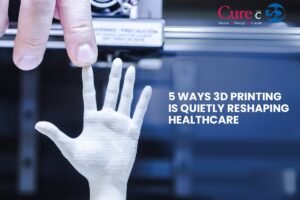
Two decades later, Chuck Hull transformed that sketch into a real object; something which you could touch & feel, a real, tangible 3D printed model. These legends opened a completely new doorway to the future.
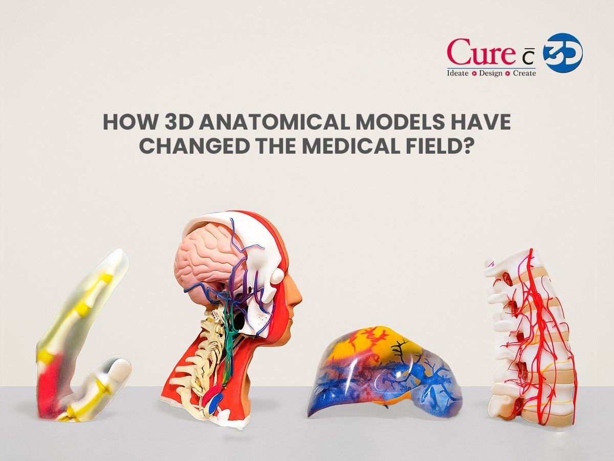
What started as a tool for engineers quickly grew beyond blueprints and prototypes. It crossed into hospitals, clinics, and medical schools, reshaping the way we heal, teach, and learn. Today, 3D anatomical models are more than just replicas of the human body. They are rehearsal stages for surgeons facing life-or-death decisions, teaching tools that bring anatomy to life for students, and bridges that help patients truly see and understand their conditions.
And this is just the beginning.
So, how exactly have 3D anatomical models transformed medicine? And what’s waiting on the horizon? Let’s dive in.
Personalized Pre-Surgical Planning
One of the clearest benefits of 3D anatomical printing lies in the surgical rehearsal it offers—something that textbooks and flat images simply can’t deliver.
Take spinal surgery: the surgical team starts by converting the patient’s CT or MRI scans into a lifelike 3D spine model, which can then be 3D printed. Holding a replica of the patient’s spine, the surgeon notes every twist, abnormal vertebra, and nerve canal. Once they run the operation through the model, the actual OR case arrives with far fewer surprises.
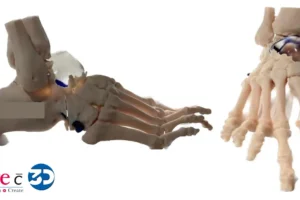
For example, before spinal surgery, a 3D model of a human spine can be printed using a patient’s CT or MRI scans. Holding a replica of the patient’s spine, the surgeon notes every twist, abnormal vertebra, and nerve canal. Once they run the operation through the model, the actual OR case arrives with far fewer surprises so, whether it’s a hand and foot anatomical model for reconstructive surgery or an eye anatomy model for ophthalmology that reproducible rehearsal delivers a greater sense of certainty and, ultimately, an improved outcome for every patient whose case arrives in the OR.
Customized Surgical Tools and Prostheses
Standard surgical tools and prostheses don’t always fit every patient. With 3D printing, surgical tools and implants can be customized to match individual anatomy. For instance, a pelvis 3D model for surgeons can be used to design implants that fit perfectly, reducing discomfort and speeding up recovery by reducing complications and ensuring higher accuracy in surgeries.
Understanding the Anatomy Like Never Before
Ask any medical student how hard it is to understand the human body’s complexities. Even though cadavers and plastic models have traditionally been used for learning, they too have their own sets of limitations, such as a limited supply of cadavers and poor preservation methods, which lead to rapid deterioration and hinder understanding.
3D printed surgical models bring anatomy to life in a far more interactive way. Imagine holding a realistic anatomical skull 3D model with intricate bone structures in your hands so that you can study from every angle. Or learning cardiology using a patient-specific heart model that shows real-life heart abnormalities, which otherwise would be difficult to study.
These lifelike models confer a level of haptic, hands-on learning that cadaver-restrictive curricula can never match. Where educators once relied on painstakingly preserved specimens, students now practice on hyper-realistic, patient-specific models, including bizarre or rare conditions. Each filament and polymer sinew can be mirrored, catalogued, and then revisited, creating an ever-evolving library of tangible anomalies that enrich every lecture.
Better Patient Education and Communication
Explaining a complex surgery to a patient can be overwhelming for both the doctor and the patient. But with patient-specific 3D printed surgical models, things have become much clearer.
For example, showing a patient their brain anatomy model or eye anatomy model can make them understand exactly what’s wrong and how the treatment will help. It builds trust, reduces fear, and helps the patients understand their health issues better & empowers them to make more informed decisions about their health.
Research and Testing Devices
Researchers often need to test medical devices, drugs, or surgical methods before they’re introduced into real practice. Using 3D anatomical models allows them to test these innovations in a controlled environment, without putting real patients at risk.
For instance, osteoporotic conditions can be studied using specialized bone models. Devices can be tested in specific pathways before being used in actual surgeries. This ensures better safety and efficiency in medical innovations.
In Forensic Science
Now, even forensic examiners rely on 3D printing. By fabricating lifelike replicas of a victim’s skull or any relevant anatomical part, specialists can break down hard cases, show what happened during an assault, save fragile reference specimens for later analysis, or demonstrate a rare anatomical variant during a trial, where flat photographs fall flat. This technology illustrates once again just how adaptable and powerful 3D models of human structure have turned out to be.
Personalized Drug 3D Printing
It may sound futuristic, but it’s already happening. 3D printing is being used to create patient-specific drug doses. Imagine a pill customised for your body’s exact needs, with precise release patterns! Even 3D printing the powdered layer to make it dissolve faster than average pills, making personalised medicine move from concept to reality.
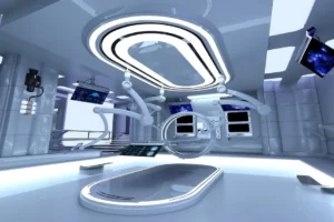
What the Future Holds: Synthetic Organs
Looking ahead, the potential of 3D anatomical models gets even more exciting. Researchers are already working on creating synthetic organs using bioprinting, which means printing human tissues with living cells.
The idea of a patient receiving a 3D-printed kidney or liver may still be in the research phase, but the progress is undeniable. In the future, this could reduce transplant waiting lists and save countless lives.
Post Processing
From affordable 3D printed surgical models to highly specialised tools like a pelvis 3D model for surgeons, 3D anatomical models are reshaping how medicine is practised. They’re making surgeries safer, education richer, research stronger, and patients more empowered.
Thanks to pioneers like Chuck Hull and years of technological advancements, what was once a dream is now a reality in hospitals worldwide. Once a futuristic idea, it has now become a lifeline, changing not only how we build, but how we heal.
References:
Narang P, Raju B, Jumah F, Konar SK, Nagaraj A, Gupta G, Nanda A. The Evolution of 3D Anatomical Models: A Brief Historical Overview. World Neurosurg. 2021 Nov;155:135-143. doi: 10.1016/j.wneu.2021.07.133. Epub 2021 Aug 5. PMID: 34363996.