
Every decision made in the operating room carries weight, sometimes, the weight of a life.
According to the World Health Organization (WHO), 1 in every 10 patients experiences harm during healthcare. Surgical errors alone account for 10% of preventable patient harm, many of which occur before or after the actual procedure.
Despite advances in medical technology, surgeons still often operate with limited spatial understanding based solely on 2D scans.
What if we could reduce that margin of error, not with better guesses, but with better guidance literally in hand?
Welcome to the New Era of Precision: 3D Printed Surgical Guides
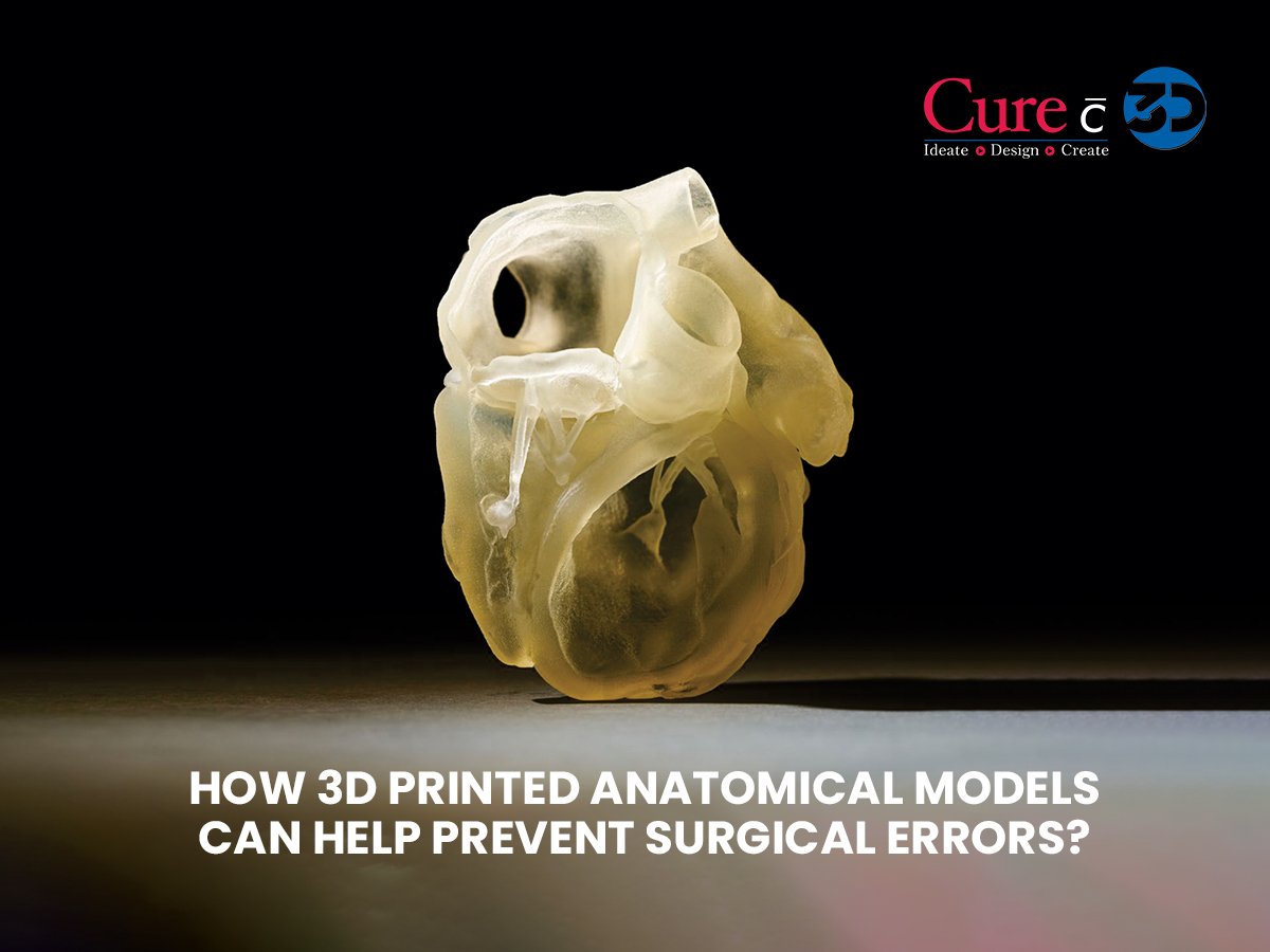
3D surgical models, a custom 3d printed anatomical models. Based on patient-specific CT or MRI scans, physical replicas of organs, joints, or bones, these models provide an incredibly accurate visual and tactile reference for surgeons before they even make the first incision. These 3D printed surgery models allow surgeons to understand the patient’s particular anatomy, anticipate complications, and practice the surgery.
How 3D Printed Models Are Made?
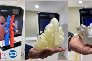
Based on the information from a CT or MRI scan, experts develop a millimetre-for-millimetre anatomical model printed with a 3D printer resembling the patient’s anatomy. Such models can range from very exact structures such as an eye anatomy model or lung anatomy model, to composite structures such as a 3D model of a knee joint or an anatomical skull 3D model.
Procedure involved:
- Collection of data in DICOM images
- 3D segmentation for conversion into STL format
- High resolution 3D printed anatomical models
The end result? A model a surgeon can hold, rotate, and even practice procedures on the 3d surgical model to get a feel of the actual surgery.
Why are Surgical Errors Still So Common?
Despite awareness of potential risks, surgical errors continue to impact patient safety. Among the 300+ million surgeries performed globally each year, many complications stem from poor pre-surgical planning, anatomical variability, or unforeseen intraoperative challenges.
3D surgical models offer:
- Pre-surgical rehearsal: Surgeons can simulate and refine their approach.
- Communication: Surgeons, patients, and even families can visualize the problem and understand the treatment better.
- Time-saving in OR: With clearer pathways planned in advance, surgeries become quicker and more efficient.
This level of preparation significantly reduces the risk of complications, re-operations, and prolonged recovery times.
Visualisation That Goes Beyond Screens
Unlike digital 3D images, tangible models bring anatomy to life. Surgeons can hold a lung anatomy model, rotate an ear anatomy model, or study the structure of a foot anatomy model from every angle. The benefits?
- Better planning: Every cut, screw, or graft is decided in advance.
- Fewer surprises: Unusual anatomy or deformities are spotted earlier.
- Improved accuracy: Enhanced orientation reduces intraoperative uncertainty.
For instance, in orthopaedics, a 3D model of a knee joint or pelvis 3D model can aid in joint replacement alignment or fracture fixation. In neurosurgery, a brain anatomy model helps map complex tumour margins while preserving healthy tissue.
Practice Before Your Actual Surgery
Athletes practice before a big game. So why not surgeons?
3D printed models allow dry runs on exact patient anatomy. Whether it’s fixing a fractured skull or navigating the spine, surgeons can rehearse on an anatomical skull 3D model or anatomical spine model as many times as needed before entering the OR.
The result?
- Shorter surgery times
- Reduced blood loss
- Lower risk of infection
- Greater confidence for the entire surgical team
Patient Education and Consent Just Got Easier
Another often-overlooked benefit is improved communication with patients and families.
Instead of explaining a procedure using abstract images, doctors can show them a hand anatomy model, an eye anatomy model, or even their own 3D printed anatomical models. This helps patients understand:
- Where the issue lies
- What the surgeon will do
- What risks and outcomes to expect
This leads to better informed consent, lower anxiety, and more trust in the care team.
3D Printed Models Applications for Every Specialty
From orthopaedics to neurosurgery, the use of 3D anatomical models spans multiple domains:
- Neurosurgery: A brain anatomy model allows for a clear visualization of the tumor location and necessary structures, which can be very essential for surgery.
- Orthopaedics: Foot anatomy models, hand anatomy models, and pelvis 3D models not only facilitate joint replacement planning but also are the trauma cases’ lifesavers.
- Cardiology: Besides an anatomical heart model, providing space for better planning in congenital defects or valve replacements is another benefit of heart models.
- Ophthalmology and ENT: As an example, the eye anatomy model, or the ear anatomy model are some of the intricate models that become the regions access plan in the case of delicate regions.
- Spine Surgery: The use of an anatomical spine model in scoliosis cases or spinal cord decompression certainly is very helpful as it offers clarity
- Pulmonology and Thoracic Surgery
Bridging Gaps in Training and Medical Education
Beyond surgery, 3D models are transforming medical education.
From resident surgeons to anatomy students, handling a 3D printed anatomical model builds spatial understanding like no textbook can. Surgeons in training can make practice cuts, simulate procedures, and gain real experience—without needing a cadaver lab.
This is particularly important in developing countries or remote areas where training infrastructure is limited.
- For trainees: Models like the anatomical skull 3D model or 3D printed pelvis provide realistic, hands-on learning opportunities that traditional atlases and VR cannot replicate.
- For patients: Explaining complex procedures becomes easier with a physical model. Patients become more informed, cooperative, and less anxious when they can see what the surgeon sees.
The result? Better communication, higher trust, and more successful outcomes.
Better Outcomes at Lower Costs
Fewer complications mean shorter hospital stays and reduced follow-up costs. Investing in 3D surgical models can:
- Reduce OR time by up to 60 minutes (in complex surgeries)
- Cut down implant mismatch and revision rates
- Improve surgical precision
- Better patient satisfaction
- Lower readmission rates
The Curewith3D Promise
At Curewith3D, we don’t just print anatomical models—we build clarity, confidence, and care. Our team works closely with surgeons to craft models that match the exact size, shape, and pathology of the patient’s anatomy. Whether it’s a pelvis 3D model, anatomical heart model, or a full 3D brain model, we deliver high-quality, medically accurate guides that change outcomes.
See Better with 3D Printed Models
Surgical errors may be a reality, but they don’t have to be inevitable. As medicine becomes more precise and personalised, 3D printed anatomical models are moving towards the forefront of that transition. By facilitating better planning, boosting accuracy, and building up patient confidence, these models are not only the means to perform surgery; they are also the means to save lives.
Ready to make your surgeries safer and smarter?Explore the power of 3D surgical guides with Curewith3D. Contact our team at curewith3d@anvka.com today!